Muscle (skeletal) of the body - definition and function
Skeletal Muscle of the Body - contractile fibers or the structures these fibers form. Muscles move the body, some under voluntary control and others reflexively. The gastrointestinal tract, genitourinary tract, and BLOOD vessels contain smooth (nonstriated) muscle, which is under involuntary control of the autonomic NERVOUS SYSTEM. The HEART contains a specialized form of muscle called myocardial, also under control of the autonomic nervous system. The bulk of the muscle tissue in the body is skeletal (striated) muscle, which responds to voluntary control through the CENTRAL NERVOUS SYSTEM.
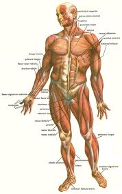
What are Skeletal Muscles and Definition
The skeletal muscles are responsible for movement and account for about 40 percent of the body’s mass. They generally appear in opposing pairs attached to BONE via tendons. When one muscle contracts, its opposing muscle relaxes. This allows smooth, balanced movement. The SKELETON provides resistance and leverage for the muscles as they contract and relax. The body contains about 650 muscles, the largest of which is the gluteus maximus (main muscle of the buttocks) and the smallest of which is the stapedius in the middle EAR (moves the stapes bone).
Movement requires interaction between neurons (NERVE cells) and muscle fibers. This interaction occurs at the neuromuscular junction, a synapse where the NEURON’s axons end (terminate) and the muscle fiber begins. When conveying a nerve impulse to activate a skeletal muscle fiber, the motor neuron releases a molecule of acetylcholine, a NEUROTRANSMITTER. The acetylcholine molecule binds with an acetylcholine receptor on the muscle fiber, forming a biochemical bridge that allows the nerve impulse to travel from the neuron to the muscle fiber. The impulse creates an action potential in the muscle fiber—a cycle of activation, discharge, and recovery—that becomes a muscle contraction.
Skeletal Muscles and Types Fibers
Skeletal muscles contain two types of fiber that dictate how rapidly and with what intensity they complete an action potential. Type 1 fibers, also called slow-twitch or red fibers (red because they have a high myoglobin content), have a slow and steady response. Type 1 fibers are in a constant state of partial contraction; they provide muscle tone and are essential for maintaining the body’s posture. Type 2 fibers, also called fast-twitch or white fibers (white because they contain very little myoglobin), have a rapid response. Type 2 fibers are responsible for muscle STRENGTH. Most skeletal muscles contain a combination of type 1 and type 2 fibers. Exercise to extend ENDURANCE increases the percentage of type 1 fibers; exercise to improve strength increases the percentage of type 2 fibers.
For further discussion of muscle within the context of the structures and functions of the musculoskeletal system, please see the overview section “The Musculoskeletal System.”
See also CELL STRUCTURE AND FUNCTION; FASCIA; LIGAMENT; PROPRIOCEPTION; TENDON.