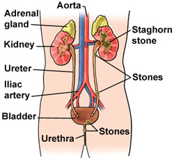
Ureter function and definition
Ureter, in a human organism, is a muscular tube, fairly thick and rigid, that leads URINE from the kidney to the urinary BLADDER. Both of the two tubular structures arise from the renal pelvis and collect the urine from the kidney’s collecting tubules. Both ureters with the inner diameter of 3 or 4 millimeters exit the kidney at the hilus. Then, the left ureter continues among the inferior VENA CAVA and the right one among the AORTA through the abdomen directly to the pelvis. At the pelvis, near to the bifurcation of the iliac arteries (ARTERY and VEIN), the ureters cross the pelvic brim, and lead into the top back of the urinary bladder. The ureter forms a short, flattened channel within the bladder wall before entering the inside of the bladder. These tunnels create a valve, also known as ureterovesical valve, helping to prevent the urine to backflow to the ureter from the bladder. Both ureters are approximately 12 inches long. However, the left one is slightly longer than the right one as the left kidney is placed about an inch higher in the abdomen. The structure of the ureters does not differ for both sexes.
The interior lining of the ureter is known as epithelial membrane and conduct of double-layered MUSCLE surrounding the urethral epithelium. The first layer surrounds the epithelium in spiral, and the other covers the ureter in a circular pattern. Both layers wave in rhythmic contractions (PERISTALSIS) in order to move the kidney closer to the urinary bladder. The outside surface of the ureter is created by a fibrous tissue.
For further discussion of the ureters within the context of the urinary system’s structure and function please see the overview section “The Urinary System.”
See also GLOMERULUS; KIDNEYS; NEPHRON; URETHRA; VESICOURETERAL REFLUX.