Electrocardiogram (ECG) diagnostic procedure
What is Electrocardiogram (ECG)
Electrocardiogram (ECG) is a noninvasive diagnostic procedure that converts the heart’s electrical activity into patterns of signals typically recorded on paper or displayed on a screen. The ECG is the cornerstone of cardiovascular diagnosis. The normal HEART generates a consistent electrical pattern; nearly anything that goes wrong with the heart shows up on an ECG. A normal heart rhythm produces five predictable fluctuations, called waves, that doctors identify by the letters P, Q, R, S, and T. The main thrust of cardiac activity, ventricular contraction, is the QRS complex.
| ECG TRACING | |
|---|---|
| P wave | sinoatrial (SA) node’s pacing impulse initiates the CARDIAC CYCLE |
| Q wave | pacing impulse arrives at the ventricular apex |
| R wave | main ventricular contraction |
| S wave | completion of ventricular contraction |
| T wave | heart’s return to readiness for the next cardiac cycle |
Reasons for Doing This Test
Electrocardiogram (ECG) is a common procedure to assess the function of the heart. It can be baseline, diagnostic, or monitoring. A baseline ECG is generally part of a ROUTINE MEDICAL EXAMINATION and establishes a record of the heart’s activity when the heart is presumably healthy. A baseline ECG provides a standard for comparison should there be cardiovascular symptoms the cardiologist needs to evaluate. The cardiologist does a diagnostic ECG to examine the heart’s electrical activity as it may correlate to symptoms the person is experiencing. The most common symptoms for which doctors conduct diagnostic ECGs are CHEST PAIN and PALPITATIONS. A monitoring ECG checks the heart’s rhythm as a means of evaluating whether medications are working effectively to treat ARRHYTHMIA or to determine whether the heart’s function is stable following heart surgery or a cardiac crisis such as HEART ATTACK.
Electrocardiogram (ECG): Preparation, Procedure, and Recovery
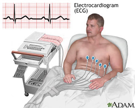
It is a good idea to avoid CAFFEINE and cigarettes for an hour or so before a scheduled ECG. Doctors generally prefer for ECG to show the heart at rest and prefer people not engage in strenuous exercise within four hours of Electrocardiogram (ECG). Otherwise, ECG requires no preparation and may take place in the doctor’s office, at a cardiovascular testing facility, or a hospital. The person lies quietly on a gurney or procedure bed and the ECG technician places about a dozen electrodes on the chest, back, arms, and legs. Talking or moving during the ECG can produce electrical “static” from the muscles. A typical ECG takes about five minutes to complete. Though the reading is immediately available, a cardiologist must interpret it and usually it takes a day for the doctor to report the results back for a routine ECG. The person may go home after the ECG recording is finished.
Variations on the standard Electrocardiogram (ECG) procedure include
- Holter monitor or ambulatory ECG, which uses a small, battery-operated unit the person wears on a shoulder strap or belt to monitor the heart’s electrical activity over a period of time, typically 24 hours (an ECG technician places the electrodes on the person’s chest and back and connects them to the unit)
- Exercise ECG, in which the person walks at varying paces on a treadmill or rides a stationary bicycle while the ECG records the heart’s changes in rhythm
- Event ECG, in which the person wears electrodes attached to a small, battery-operated unit that the person turns on during a cardiac event such as palpitations
Electrocardiogram (ECG): Risks and Complications
There are no risks or complications associated with ECG. Sometimes the ECG technician must shave a small area of SKIN to allow good electrode contact. Some people who are highly sensitive to adhesive may have a slight skin reaction to the adhesive pad that holds the electrode in place. And some people quickly chill when lying on the procedure table; the ECG technician can cover the person with a warm blanket for improved comfort and to prevent shivering, which can distort the ECG reading. ECG only detects and records the electrical activity of the heart; it does not send any electrical impulses to the heart.
See also AUTOMATED EXTERNAL DEFIBRILLATOR (AED); CARDIOVERSION; DEFIBRILLATION; ECHOCARDIOGRAM.