Angiogram - procedure and risks
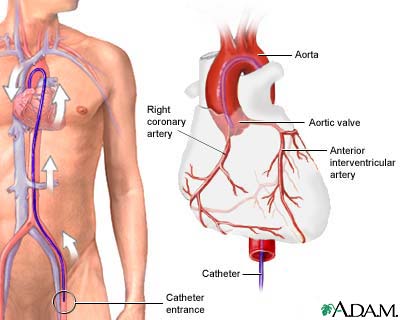
Angiogram is a diagnostic test to visualize BLOOD vessels. The test is an angiography; the result is an angiogram. The cardiologist or vascular specialist injects dye into the relevant blood vessels to assess the flow of blood through them, observing the flow via FLUOROSCOPY (moving X-rays). Angiography is useful for diagnosing PERIPHERAL VASCULAR DISEASE (PVD), CORONARY ARTERY DISEASE (CAD), VENOUS INSUFFICIENCY, and DEEP VEIN THROMBOSIS (DVT). The cardiologist does angiography of the HEART during CARDIAC CATHETERIZATION. The risks of angiography include bleeding or INFECTION at the injection site and reaction to the dye. With the precision and availability of noninvasive imaging technology such as COMPUTED TOMOGRAPHY (CT) SCAN and MAGNETIC RESONANCE IMAGING (MRI), doctors use noncardiac angiography (angiography of peripheral blood vessels such as in the legs) primarily when the diagnosis or extent of blockage remains uncertain or before surgery to correct blockages.
See also ANGIOPLASTY; CORONARY ARTERY BYPASS GRAFT (CABG).